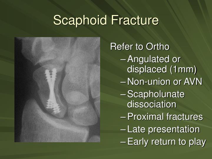
MRI imaging can be helpful as it can image soft tissues and detect tears of both intrinsic and extrinsic ligaments. Both of these imaging studies will show fractures earlier-if there is no displacement versus plain radiographs. At times, enhanced imaging including CT scans or MRI is a helpful adjuvant. These films may be repeated in two weeks to assure there is not an occult scaphoid fracture. Typically routine x-rays are sufficient, although they may be taken from many angles.Īs noted above, initial radiographs for scaphoid fractures may be negative. X-rays help delineate the type of fracture, displacement and if the fracture extends within a joint.

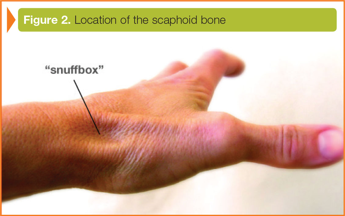
Although x-rays do not image soft tissues, such as ligaments, they are the first line evaluation in looking for fractures of the limb. X-rays of the wrist are obtained and if there is suspicion of injury to the hand, elbow or shoulder these may be obtained as well. Tenderness to palpation is typically elicited over the base of the thumb-often called the anatomic snuff box. Any deformity of the hand or wrist is also noted. Swelling from the sprains may cause compression of vascular structures leading to changes in blood supply to the hand. The most common nerve injured is the median nerve, resulting in numbness in the radial three digits of the hand. Sometimes patients with scaphoid fractures may have injured the nerves associated with the hand. Although unlikely, injuries to the adjacent shoulder and elbow are determined via checking for pain and motion.Īn examination of the sensation to the hand is performed. A physical exam centers on the injured limb. Hand Surgeon ExaminationĪfter taking note of the symptoms, the surgeon inquiries regarding any pertinent family or medical history. These symptoms are typically worse with gripping and wrist motion. Typically the injury occurs after a traumatic event-like a fall. Patients with a scaphoid fracture typically complain of pain, bruising, and swelling. The extrinsic ligaments, note these ligaments may span 2 or more joints. These ligaments are not as dense or as strong as the intrinsic ligaments. The next layer of ligaments lying more superficial than the intrinsic ligaments are the extrinsic ligaments. The most important intrinsic ligaments are the SL (scapholunate) and LT (lunotriquetral) The intrinsic ligaments of the wrist from the top right A and bottom B. Since these ligaments are inside the wrist-they are called intrinsic ligaments.
SCAPHOID PAIN BUT NO FRACTURE SERIES
These bones are interconnected with a series of ligaments. The eight bones of the right wrist (carpus) viewed from the front.

The wrist is made up of eight carpal bones connecting the forearm to the hand. The ligaments of the wrist are external to the wrist and internal to the wrist. The scaphoid has a particularly poor blood supply and gaining healing of this bone can be difficult-complications with treatment and healing are common. Treatment of these fractures spans from nonoperative treatment in a cast or brace to surgical management. These injuries often masquerade as wrist sprains-and initial radiographs may not reveal the fracture. These fractures are often associated with tenderness on the top of the wrist. The scaphoid (navicular) is one of the proximal carpal bones and may be injured in a fall. A Patient’s Guide to Scaphoid (Navicular) Fracture with Animated Surgical Video Introduction


 0 kommentar(er)
